Cross Section Of A Bone Diagram : Bone Structure Anatomy Explained What Is Bone Marrow : In the last decade, considerable technological improvements have been made to repair damaged bones and tissue, such as bone cross sections with implants for microscopic examinations.
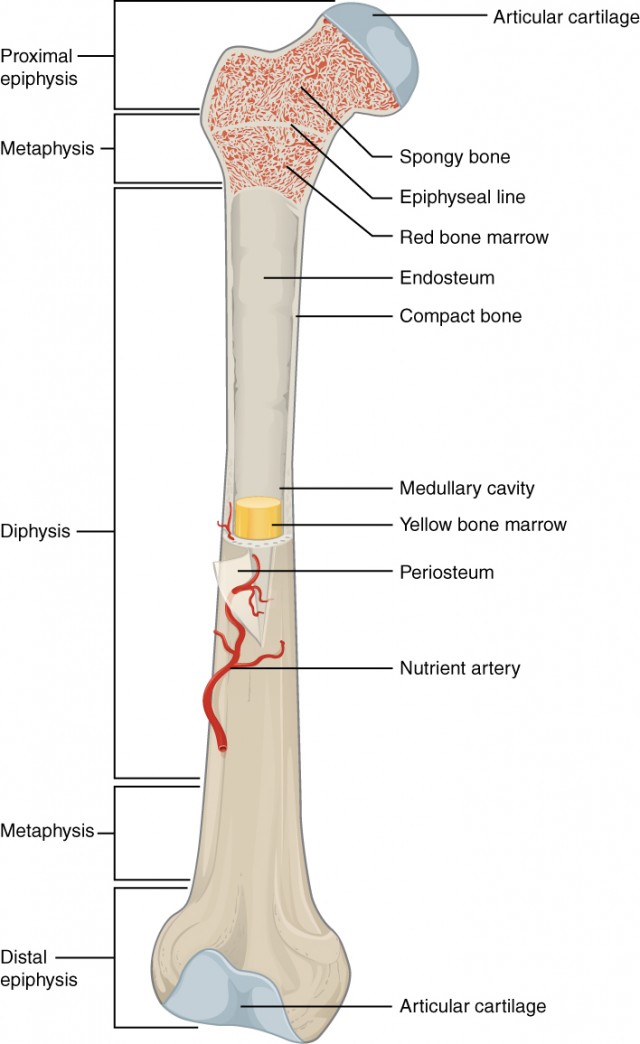
Cross Section Of A Bone Diagram : Bone Structure Anatomy Explained What Is Bone Marrow : In the last decade, considerable technological improvements have been made to repair damaged bones and tissue, such as bone cross sections with implants for microscopic examinations.. Jump to navigation jump to search. Bone is found in the shafts of long bone and consists of various cylindrical units named as haversian system 47. Diagram with articular cartilage, marrow, spongy bone, medullary cavity, endosteum, diaphysis, and periosteum. Whereas a long bone has only one layer of compact bone (see fig 1). Bone marrow is the soft, highly vascular and flexible connective tissue within bone cavities which serve as the primary site of new blood cell production or bone marrow is the primary source of pluripotent stem cells that give rise to all hemopoietic cells (blood cells) including lymphocytes.
A cross section of a human long bone. Whereas a long bone has only one layer of compact bone (see fig 1). Diagram with articular cartilage, marrow, medullary cavity and periosteum. Cross‐sectional area is derived from the integral of the bone mass profile across the narrow region. As with other tools applied to petroleum development.
Bone Marrow And The Immune System from assets.aboutkidshealth.ca The large dark spots are passages for blood vessels and nerves. Explaned distal and proximal epiphysis. Vector illustration scheme of bone cross section. The centroidal distance, c, is the distance from the centroid of a cross section to the extreme fiber. The well known 67 nm periodic pattern results from the. Compact bone cross section courtesy: Diagram with articular cartilage, marrow, spongy bone, medullary cavity, endosteum, diaphysis, and periosteum. can be used for personal and commercial purposes. This simply involves placing a section of the bone on the microscope stage and viewing the.
Bone marrow is the soft, highly vascular and flexible connective tissue within bone cavities which serve as the primary site of new blood cell production or bone marrow is the primary source of pluripotent stem cells that give rise to all hemopoietic cells (blood cells) including lymphocytes.
Cord spinal cross section spine cervical diagram education science anatomical anatomy atlas back body bone care column disc disease foramen fracture grey health healthcare healthy human illustration infographic injury matter medical nerve nervous pain part physiology poster process skeletal skeleton. Bone tissue cross section diagram human oasissolutions co. They build the entire picture, improve your understanding, consolidate the information and facilitate recall. Geological cross sections are graphical representations of vertical slices through the earth used to clarify or interpret geological relationships with or without accompanying maps. This page discusses the calculation of cross section properties relevant to structural analysis, including centroid, moment of inertia, section modulus, and parallel axis theorem. Cross section of bone diagram. We can see there are two layers of compact bone here. Explaned distal and proximal epiphysis. Diagram with articular cartilage, marrow, spongy bone, medullary cavity, endosteum, diaphysis, and periosteum. can be used for personal and commercial purposes. Vector illustration scheme of bone cross section. Compact bone cross section courtesy: Cross‐sectional area is derived from the integral of the bone mass profile across the narrow region. Explaned distal and proximal epiphysis.
Explaned distal and proximal epiphysis. Cross section diagram of human bone, bone, cross section diagram this slide contained a cross section of a very small bone, and you are looking at the entire thickness of the shaft of the bone. Looking at a bone in cross section, there are several distinct layered regions that make up a bone. The well known 67 nm periodic pattern results from the. (micrograph provided by the regents of university of michigan.
Bone Structure Anatomy And Physiology I from s3-us-west-2.amazonaws.com Cord spinal cross section spine cervical diagram education science anatomical anatomy atlas back body bone care column disc disease foramen fracture grey health healthcare healthy human illustration infographic injury matter medical nerve nervous pain part physiology poster process skeletal skeleton. As shown in figure 2. A diagram of the relative position of the bone, cartilage, and synovial membrane. The periosteum contains many strong collagen fibers that are used to firmly anchor. Diagram with articular cartilage, marrow, medullary cavity and periosteum. Explaned distal and proximal epiphysis. A cross section of a human long bone. This page discusses the calculation of cross section properties relevant to structural analysis, including centroid, moment of inertia, section modulus, and parallel axis theorem.
It seems confusing and misleading.
Whereas a long bone has only one layer of compact bone (see fig 1). (micrograph provided by the regents of university of michigan. Compact bone cross section courtesy: I am not an expert on this subject, so i was wondering if i don't like way you've shown the cartilage. We can see there are two layers of compact bone here. The outside of a bone is covered in a thin layer of dense irregular connective tissue called the periosteum. The well known 67 nm periodic pattern results from the. Bone is found in the shafts of long bone and consists of various cylindrical units named as haversian system 47. As the names suggest compact bone looks compact and the spongy bone looks like skull bone is a flat bone. Cross‐sectional area is derived from the integral of the bone mass profile across the narrow region. Cord spinal cross section spine cervical diagram education science anatomical anatomy atlas back body bone care column disc disease foramen fracture grey health healthcare healthy human illustration infographic injury matter medical nerve nervous pain part physiology poster process skeletal skeleton. The cross section of a rectangular pyramid is a rectangle. Diagram with articular cartilage, marrow, medullary cavity and periosteum.
The spinal cord is elliptical in cross section being spinal cord crosssection images stock photos vectors shutterstock. Cord spinal cross section spine cervical diagram education science anatomical anatomy atlas back body bone care column disc disease foramen fracture grey health healthcare healthy human illustration infographic injury matter medical nerve nervous pain part physiology poster process skeletal skeleton. Please like this video share with all the learners comment your opinion and subscribe to support my channel this is a small step to teach what i know to. As the names suggest compact bone looks compact and the spongy bone looks like skull bone is a flat bone. Bone tissue cross section diagram human oasissolutions co.
Axilla Cross Section Cadaveric Anatomy Boundaries And Contents Youtube from i.ytimg.com The cross section of a rectangular pyramid is a rectangle. Fermur bone with labels and diagram. Medically reviewed by the healthline medical network — written by the healthline editorial team — updated on january 20, 2018. (left) a schematic diagram illustrating the assembly of collagen fibrils and fibers and bone mineral crystals. Vector illustration scheme of bone cross section. The outside of a bone is covered in a thin layer of dense irregular connective tissue called the periosteum. The spinal cord is elliptical in cross section being spinal cord crosssection images stock photos vectors shutterstock. (micrograph provided by the regents of university of michigan.
Explaned distal and proximal epiphysis.
As shown in figure 2. The cross section of a rectangular pyramid is a rectangle. Explaned distal and proximal epiphysis. Bone tissue cross section diagram human oasissolutions co. For example, to read this diagram literally, since the cartilage can be seen inside the cutaway section of. The cross section of this circular cylinder is a circle. A diagram of the relative position of the bone, cartilage, and synovial membrane. Bone is found in the shafts of long bone and consists of various cylindrical units named as haversian system 47. Vector illustration scheme of bone cross section. This simply involves placing a section of the bone on the microscope stage and viewing the. Bone is hard and many of its functions depend on that characteristic hardness. Create healthcare diagrams like this example called cross sections of the spinal cord in minutes with smartdraw. Jump to navigation jump to search.
Explaned distal and proximal epiphysis cross section of a bone. A diagram of the relative position of the bone, cartilage, and synovial membrane.


0 Komentar